
ISO 10993 Biocompatibility Cytotoxicity Test
Cytotoxicity Test Introduction
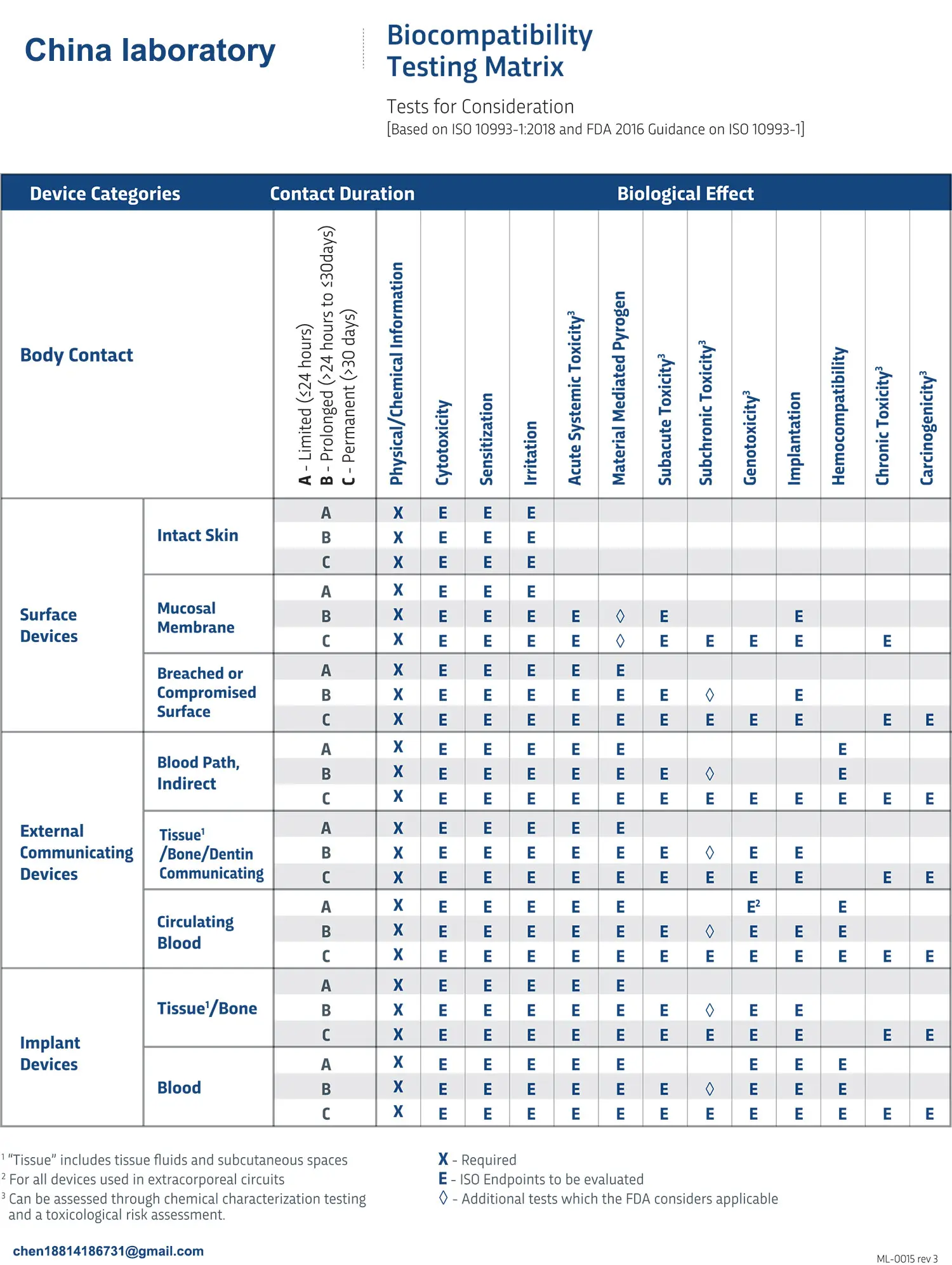
Cytotoxicity evaluates the toxic responses of potential bioactive substances in material extracts to cultured mammalian cells ().
The principle is that the yellow aqueous solution MTT is metabolically reduced in living cells to generate insoluble formazan. The number of living cells is related to the colorimetry of the formazan after it is dissolved in alcohol and measured by a photometer.
Cytotoxicity Test standards
2.1 GB/T 16886.5-2022 Biological evaluation of medical devices Part 5: In vitro cytotoxicity test;
2.2 ISO 10993-5:2009 Biological evaluation of medical devices—Part 6: Tests for in vitro cytotoxicity。
3. Test system
L929 cells.
4. Preparation of test samples
The extract was prepared according to GB/T 16886.12.
5. Test steps
5.1 Cell culture preparation
The cultured cells were removed from the culture flask by enzymatic digestion (trypsin/EDTA) and the cell suspension was centrifuged at 200 g for 3 min. The cell suspension was resuspended in culture medium to adjust the density to 1 × 10⁵ cells/mL and 100 μL of culture medium was added to the peripheral wells of a 96-well tissue culture microtiter plate (= 1 × 10⁴ cells/well) using a multichannel pipette.
The cells were incubated for 24 h to form a semi-confluent monolayer. The culture was incubated in a CO2 incubator (37°C ± 1°C, humidity > 90%) with a 5 ± 1% atmosphere for (24 ± 2) h to allow the cells to grow to form a nearly confluent monolayer.
5.2 Contact training
After 24 hours of incubation, aspirate the culture medium from the plate wells and add 100 μL of the test sample extract, control sample extract, blank control solution and positive control solution. All doses need to be done in at least six parallels. Add the blank to the left (column 2) and right (column 11) sides of the 96-well plate. Then incubate for 24 hours ± 2 hours at (37 ± 1) ° C, 5 ± 1% CO2, and humidity > 90%.
5.3 Observation
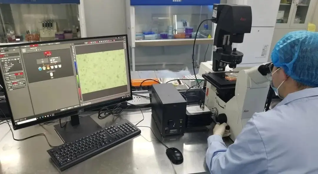
After 24 hours of incubation, examine each plate under a phase contrast microscope to determine the systematic errors in cell seeding and the growth characteristics of the control and test cells. Record changes in cell morphology caused by the cytotoxic effects of the test sample extracts, but these records are not used in any quantitative determination of cytotoxicity. Poor growth characteristics of the control cells may indicate experimental errors and the test may be abandoned as a result.
5.4 Add MTT
After checking the cell growth in the plate wells, carefully remove the culture medium from the plate wells, add 50 μL of MTT solution (prepared to 1 mg/mL with complete MEM culture medium without phenol red) to each test plate well, and culture at (37 ± 1) ℃ for (2 ± 0.2) h. Discard the MTT solution, add 100 μL of isopropanol to each well to dissolve the formazan, shake the plate, and then measure the absorbance value of each well at a wavelength of 570 nm (reference wavelength 650 nm).
5.4 Data Analysis
The reduction in the number of viable cells results in a decrease in metabolic activity in the sample, which is directly related to the amount of blue-violet formazan produced. The reduction in cell viability of the test sample compared to the blank control ( cells directly in contact with the extraction medium ) can be calcULated using the following formula:
MICrosoft YaHei", "Microsoft YaHei UI", "WenQuanYi Micro Hei", "sans-serif", 宋体; font-optical-sizing: inherit; font-kerning: inherit; font-feature-settings: inherit; font-variation-settings: inherit; margin-top: 0px; margin-bottom: 0px; padding: 0px; color: rgb(51, 51, 51); box-sizing: border-box; text-wrap: wrap; background-color: rgb(255, 255, 255);">
Where: OD570e---the average absorbance value measuRED for the test sample, positive control and negative control groups.
OD570b---average absorbance value of the blank control group (cells directly in contact with the extraction medium).
When the survival rate is low, the potential cytotoxicity of the test sample is high. If the survival rate decreases by less than 70% of the blank, the fish oil is potentially cytotoxic. The survival rate of the 50% extract of the test sample is the same or higher, otherwise the test should be repeated.
Email:hello@jjrlab.com
Write your message here and send it to us
 EMC Item – Introduction to Radiated Emission Test
 IEC 62471 Photobiological Safety of Lamps and Lamp
 New European Toy Standard EN 71-1:2026
 EN71 Series Standards Compliance February 13, 2026
 European Toy Safety Standard EN 71-20:2025
 EN 18031 Certification for Connected Devices on Am
 Compliance Guide for Portable Batteries on Amazon
 2026 EU SVHC Candidate List (253 Substances)
Leave us a message
24-hour online customer service at any time to respond, so that you worry!
