


Cytotoxicity Test - CMA+CNAS Accredited Laboratory
A cytotoxicity test refers to evaluating the toxic effects of various chemical substances used as raw materials in developing medical devices, some of which may have certain toxic effects in the body. The cytotoxicity test assesses the toxic effects eluted from medical devices and the cell death and morphological changes caused by the chemical substances used as raw materials. It is an essential test for all medical devices.
")
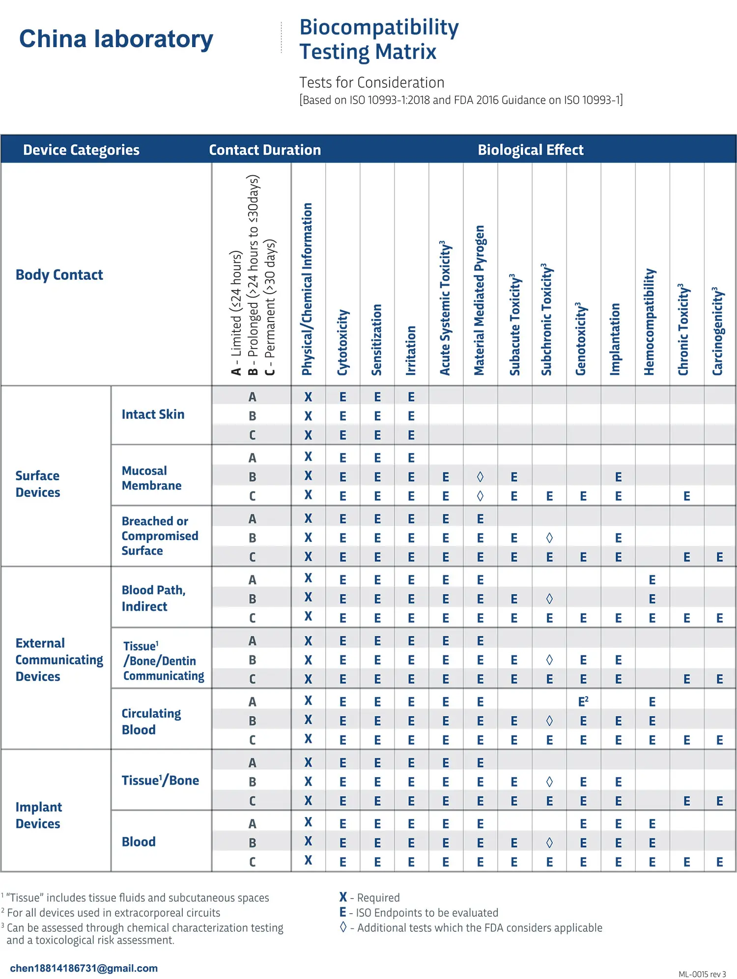
China JJR Laboratory is a CMA+CNAS accredited laboratory providing iso 10993 cytotoxicity test services. Below is a detailed explanation of cytotoxicity testing methods:
1. Colony Formation Assay
Objective: Evaluate the cytotoxic effect by detecting the colony formation rate in culture medium extracts of the tested medical device.
Cells Used: Chinese hamster V79 strain, Mouse L929 strain, Mouse BALB/c 3T3 strain
Overview: This is a highly sensitive in vitro test system that can replace animal-based irritation and acute toxicity tests. The colony formation test is conducted in the extract, where the test subject is incubated with the culture medium in an amount proportional to its surface area (or weight). Control materials with known cytotoxic effects are also tested for comparison to infer in vivo cytotoxic effects. This test is essential for GLP (Good Laboratory Practice) in medical devices.
2. Direct Contact Test
Objective: Conduct a colony formation assay on the test subject to evaluate cytotoxicity.
Cells Used: Chinese hamster V79 strain
Overview: The tested medical device is cut to a specific size, immersed in culture medium, and cells are directly seeded onto it to detect colony formation rates. Since cells are more sensitive in this method, it allows for toxicity evaluation.
3. Tissue Culture (TC) Insert Test
Objective: Conduct a colony formation test on TC inserts placed inside an incubator containing the test subject to assess cytotoxicity.
Cells Used: Chinese hamster V79 strain
Overview: This method serves as a supplement when the shape or surface of the test subject is unsuitable for direct contact testing. The tested material is cut to a specific size and placed in close contact with the bottom of a culture incubator containing culture medium. A TC insert is then placed on top. Cells are seeded onto the TC insert, and a colony formation test is conducted while coexisting with the test subject.
4. Elution Test
Objective: Evaluate cytotoxic effects by treating monolayer-cultured cells with culture medium extracts from the tested medical device.
Cells Used: Chinese hamster V79 strain, Mouse L929 strain, Mouse BALB/c 3T3 strain, Human MRC-5 strain
Overview: Similar to the colony formation assay using culture medium extracts, the test involves preparing culture medium extracts of the test subject and applying them to subconfluent monolayers. Cultured cells on plates are treated with the extract to assess growth rate. Cell proliferation is measured by staining with crystal violet and using a monolayer cell density meter (monocerator).
5. Direct Contact Test
The test subject is placed in an incubator containing monolayer-cultured target cells, and cytotoxicity is evaluated under direct contact conditions.
Overview: The test subject is placed on cells cultured in a monolayer at the subconfluent stage. The inhibition of cell proliferation around and beneath the test subject is observed to assess cytotoxic effects. Cytotoxicity is evaluated using a five-point scale based on morphological observations of the cells.
6. Neutral Red (NR) Assay
Objective: Assess the cytotoxic effects of target chemical substances.
Cells Used: Mouse BALB/c 3T3 strain
Overview: This method utilizes the ability of viable cells to absorb neutral red dye. BALB/c 3T3 cells cultured in plates are treated with chemical substances, and neutral red is subsequently extracted to measure absorbance. This method is commonly used for evaluating cytotoxic effects. Additionally, it is applied to assess photo-cytotoxicity by treating chemical substances under light exposure conditions.
Email:hello@jjrlab.com
Write your message here and send it to us
 Toothbrush FDA Certification Testing
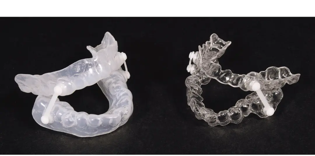 Snoring Device FDA 510k Standard Testing
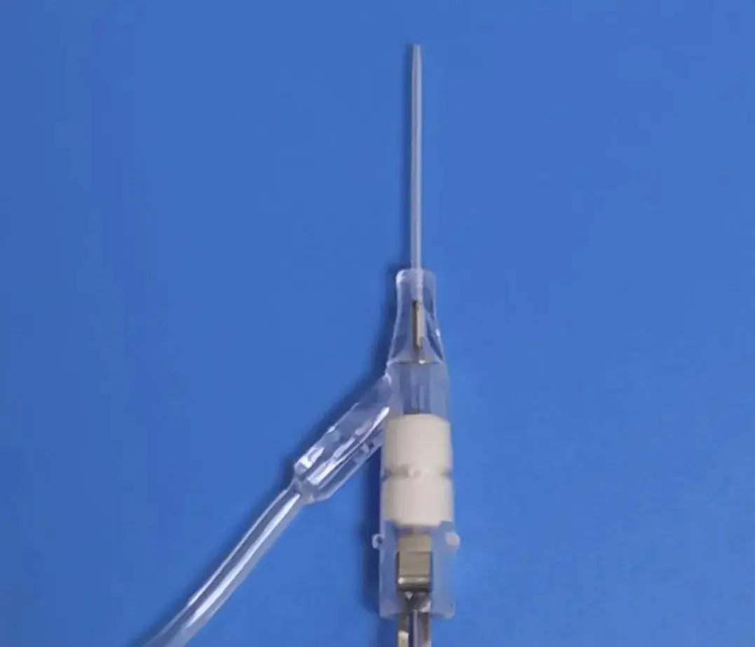 Single Use Intravenous Catheter Certification Test
 Silicone Material Product Compliance Certification
 What to Do If Cytotoxicity Test Results Are Positi
 ISO 10993:5 Cytotoxicity Testing Methods
 FDA ISO 10993-1 Biocompatibility Evaluation Guidel
In Vitro Cytotoxicity Testing for Medical Devices
FDA ISO 10993-1 Biocompatibility Evaluation Guidel
In Vitro Cytotoxicity Testing for Medical Devices
Leave us a message
24-hour online customer service at any time to respond, so that you worry!