


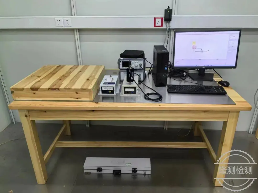
Medical Device Subchronic Systemic Toxicity Testing Laboratory
1. Definition
Adverse effects that occur at a certain stage in the animal's life span after repeated or continuous exposure to the test sample. Subchronic toxicity studies in rodents are generally 90 days, and in other species of animals, the exposure period does not exceed 10% of their life span. Subchronic intravenous studies generally stipulate that the exposure time is 14 days to 28 days.
")
2. Test basis
This test is based on ISO 10993-11 Appendix H.
3. Test system
Rodents (rats, mice) are generally used. The use of non-rodent animals should be scientifically justified.
4. Test steps
4.1 Sample preparation
Before each test, prepare the sample polar extract and non-polar extract according to iso 10993-11
4.2 Animal testing
Twenty-four animals were randomly divided into an extract group and a control group, with half of each group consisting of males and half females.
Starting from day 0, the rats were injected for 14 consecutive days. The rats were weighed and recorded before each injection. The polar extract was injected once into the tail vein of the sample extract group, and the polar control solution was injected once into the tail vein of the control group. The non-polar extract and non-polar control solution were injected intraperitoneally once on days 1, 4, 7, 10 and 13. The rats were euthanized and dissected 24 hours after the last injection. See Table 1.
The animals were fasted for 12 h before dissection, and their fasting body weights were measured. After anesthesia, blood was taken from the heart, and various indicators of hematology and blood biochemistry were measured respectively. After the animals were killed by bleeding from the abdominal aorta, each tissue and organ was dissected and observed for abnormalities, the wet weight of each organ was weighed, and the weight of each tissue and organ was calculated. Organ coefficient. After each organ was soaked in 10% formalin fixative for 72 hours, routine histopathological examination was performed.
Table 1. Recommended dosage parameters for dual-route administration
Number of animals/sex/groupa | way | volume mL/kgb | Dosing frequency Number of research days d | Injection speed ml/min |
6 | vein | 10 | Inject every day for 14 consecutive days | ≤2 |
Abdominal cavity | 5c | 1,4,7,10,13 | slow | |
a. Animals in the solvent control group (6 animals/sex) need to be given the same medication. b. Recommended injection dose volume. c. Cottonseed oil is the best choice. d. The number of dosing days starts from day 0. | ||||
4.3 Animal weight
Animal body weights were measured immediately before sample administration, and each week after the first exposure (if required during the trial period) and at the end of the trial.
4.4 Clinical Observation
The observation frequency and interval should be determined based on the nature and severity of the toxic reaction, response speed and recovery period. In the early stages of a trial, the number of observations may need to be increased. The timing and duration of signs of toxicity and the time to death of the animal are important, especially if there are signs of delayed adverse clinical symptoms or death. To avoid unnecessary suffering, animals should be subjected to humane endpoints specified in national or international animal welfare guidelines. Basic clinical observations after exposure to the test sample should take into account the peak of the expected effect.
Observations should be recorded systematically and records of observations for each animal should be kept. Survival and significant clinical responses observed should be recorded at least once a day using the laboratory's common clinical response terms. Long-term repeated exposure studies should record animal morbidity and mortality at least twice a day. Longer-term repeated exposure studies may consider recording a wider range of adverse clinical symptoms at least once a week.
Observe and record the general status of the animals in the test group and the control group daily, such as skin, coat, eye and mucous membrane changes, as well as respiratory, circulatory, autonomic and central nervous system and behavioral performance. Report using the descriptors given in Table 2.
The number of dead animals was recorded and autopsied promptly, and moribund animals were isolated and killed promptly.
Typical ophthalmic examinations use an ophthalmoscope or other similar suitable equipment and should be performed before exposure to the test substance and during the test. It is best to examine all animals, at least the high-dose and control groups. If ocular changes are detected, all animals should be examined. Abnormal inspection should be documented and demonstrated.
Table 2. Common clinical symptoms and observation items
Clinical Observation | Watch for symptoms | Systems involved |
breathe | Dyspnea (abdominal breathing, wheezing), apnea, cyanosis, tachypnea, nasal discharge | Central nervous system (CNS), lungs, heart |
Muscle movement | Decreased or worsened drowsiness, lack of righting, lack of sensation, general stiffness, ataxia, abnormal movements, proneness, tremor, fasciculations | CNS, somatomuscular, sensory, neuromuscular, autonomy |
Spasticity | Clonic, tonic, forced clonus, syncope, opisthotonus | CNS, neuromuscular, autonomic, respiratory |
reflection | Cornea, correction, stretch, light response, startle reflex | CNS, sensation, autonomy, neuromuscular |
Eye symptoms | Tearing, miosis/dilation, exophthalmos, upper face droop, opacity, iritis, conjunctivitis, haematophora, nictitating membrane relaxation. | autonomy, excitement |
cardiovascular symptoms | Bradycardia, tachycardia, arrhythmia, vasodilation, vasoconstriction | CNS, autonomy, heart, lungs |
Drooling | excessive | Autonomy |
Piloerection | Rough coat | Autonomy |
loss of pain | Reduced response | CNS, feeling |
Muscle status | hypotonia, hypertonia | Autonomy |
Gastrointestinal | Loose stools, diarrhea, vomiting, polyuria, runny nose | CNS, autonomic, sensory, gastrointestinal motility, kidney |
skin | Edema, erythema | Tissue damage, irritation |
4.5 Pathology
4.5.1 Clinical pathological examination
4.5.1.1 Conduct hematological examinations during the test exposure period and/or at the end of the exposure, including: coagulation (PT, APTT), hemoglobin concentration, hematocrit, platelet count, red blood cell count, white blood cell count and white blood cell differential count, etc.;
4.5.1.2 Conduct clinical blood biochemistry examinations during the experimental exposure period and/or at the end of the exposure, including electrolyte balance, carbohydrate metabolism, and liver and kidney function. Due to their specific mode of action, some test articles may also need to measure the following: albumin, ALP, ALT, AST, calcium, chloride, cholesterol, creatinine, CGT, glucose, inorganic phosphorus, potassium, sodium, total bilirubin, total protein, triglycerides, urine nitrogen, various enzymes, etc. When evaluating immunotoxicity, the total immunoglobulin level may be considered.
Urine testing is not a routine test and is only considered when toxic reactions are expected or observed. Urine is collected regularly during the last week of experimental contact. The following tests are recommended: appearance, bilirubin, urine sugar, ketone bodies, occult blood, protein, sediment, specific gravity or osmotic pressure, and urine volume.
4.5.2 Gross pathological examination
4.5.2.1 At the end of the test contact, all test animals will be sacrificed painlessly and a gross autopsy will be performed, including body surface and body surface openings, head, chest (abdominal) cavity and internal organs, etc.
4.5.2.2 Weigh the wet weight of the adrenal gland, brain, epididymis, heart, kidney, liver, ovary, spleen, testicle, thymus and uterus of the test animal as soon as possible after removal to prevent dryness and resulting weight loss.
4.5.2.3 Organs or tissues that require further histopathological examination shall be selected based on the expected route of action of the test article, as well as the target organs of the test article, organs and tissues showing signs of gross damage or size changes, and tissues at the site of test article implantation, and removed and placed in an appropriate fixative (10% formaldehyde fixative).
The organs that should be removed are: adrenal gland*, aorta, bone marrow (femur, ribs or sternum), brain* (representative parts, including cerebrum, cerebellum and pons), cecum, colon, duodenum, epididymis*, Esophagus, eyes, gallbladder (if any), heart*, ileum, jejunum, kidney*, liver*, lungs and bronchi (expanded and preserved by fixative), lymph nodes (local lymph nodes cover the contact site, distal lymph nodes cover the whole body function), mammary gland (female), muscle (skeletal), turbinate (for inhalation studies), nerve (sciatic or tibial nerve, preferably close to muscle), ovary*, pancreas, parathyroid gland, pituitary gland, prostate, rectum , salivary glands, seminal vesicles, skin, spinal cord, spleen*, sternum, stomach, testicles*, glands*, thyroid, trachea, bladder, uterus* (including cervix and fallopian tube), vagina.
4.5.2.4 Histopathological examination
Many medical devices use common materials that differ only in the amount or type of chemical additives, processing or sterilization methods. Based on the toxicological risk assessment of the device's extractables/leachables, if it is determined that a biocompatibility/safety evaluation is required for its potential systemic toxic effects, a simplified histopathological evaluation may be considered. In this model, a tiered approach is used to examine a limited number of target organs/tissues.
Qualified histopathological analysis should include all layer I organs/tissues listed in Table 3. If abnormal or suspicious results are observed in Tier I tissue or concurrent clinical pathology (clinical biochemistry and hematology), perform Tier II analysis (examination of all organs/tissues listed above).
Table 3. List of defined histopathological organs
Organ systems | Organs/tissues in each system (species if applicable) | Tier I Organization |
cycle | Heart, arteries, veins, capillaries, blood | heart |
Digestion | Mouth, salivary glands, esophagus, liver, stomach, gallbladder, pancreas, intestines (duodenum, transverse colon, ascending colon) Intestine, descending colon, ileum, jejunum, cecum, sigmoid colon), rectum, anus | liver |
endocrine | Hypothalamus, pituitary gland, thyroid, parathyroid glands, adrenal glands, pineal gland, pancreas | Adrenal glands |
excretion | Kidneys, ureters, bladder, urethra, skin, lungs, rectum | kidney |
Body | Skin, subcutaneous tissue, hair, nails | skin |
lymph | Lymph nodes, tonsils, adenoids, thymus, spleen | spleen |
nerve | Brain, spinal cord, nerves, peripheral nerves, eyes, ears | brain |
Reproduction | Ovary, fallopian tube, uterus, vagina, breast, testis, vas deferens, seminal vesicle, prostate, penis | ovaries, testicles |
breathe | Nose, nasal cavity, pharynx, larynx, trachea, bronchi, lungs | Lungs, bronchi |
skeleton | Femur, humerus, radius, ulna, skull, sternum, clavicle, fibula, tibia, spine, scapula, pelvis, coccyx, phalanges, bone marrow, cartilage, ligaments | femur or sternum |
Hematopoiesis | marrow | femur, ribs, or sternum |
other | Gross injury including the tested area | Organ or tissue observed |
5. Result evaluation
5.1 The table gives various test data, including the number of animals in each test group at the beginning of the test, the number of animals showing signs of damage, the type of damage, and the percentage of animals with each type of damage. Statistical methods should be used to analyze and evaluate the data, but biological relevance must first be considered.
5.2 Findings from repeated exposure studies should be evaluated in the context of previous findings, toxicological considerations, and autopsy and histopathology findings.
5.3 Analyze and evaluate the correlation between the experimental exposure dose and the toxic reactions, the incidence and severity of abnormal reactions, including behavioral and clinical abnormalities, gross lesions, microscopic changes, identification of target organs, mortality, and any other significant general and specific reactions.
Email:hello@jjrlab.com
Write your message here and send it to us
 Guide to Compliance Certification for Projector Ex
 US Clothing 16 CFR 1610 Flammability Test + GCC +
 Amazon US Button Cell Battery UL 4200A + 16 CFR 12
 AliExpress US Site: Wireless Products Must Have FC
 EU CE LVD, EMC, and RED Directives for Electrical
 How to get a UL Test Certificate
 How to get a DOE Certificate of Compliance
 California 65 Compliance Certificate
Leave us a message
24-hour online customer service at any time to respond, so that you worry!